Light microscopes
Greg Foot explains the main differences between light and electron microscopes
We need microscopes to study most cells. Microscopes are used to produce magnified images. There are two main types of microscope:
- light microscopes are used to study living cells and for regular use when relatively low The amount that an image of something is scaled up when viewed through a microscope. and The fineness of detail that can be seen in an image - the higher the resolution of an image, the more detail it holds. In computing terms, resolution is measured in dots per inch (dpi). is enough
- electron microscopes provide higher magnifications and higher resolution images but cannot be used to view living cells
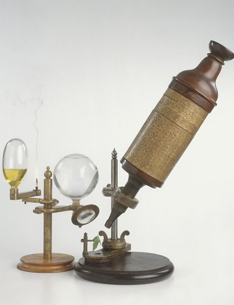
How have light microscopes developed?
Glass was developed by the Romans in the first century. Since then, scientists have been trying to magnify objects. No-one knows who first invented the microscope, but there have been key stages in their development:
1590s: Dutch spectacle maker Janssen experimented with putting lenses in tubes. He made the first compound microscope. None of these microscopes have survived, but they are thought to have magnified from ×3 to ×9.
1650s: British scientist, Robert Hooke (also famous for his law of elasticity in Physics) observed and drew cells using a A microscope in which the lens is close to the sample being magnified..
Late 1600s: Dutch scientist Antonie van Leeuwenhoek constructed a microscope with a single spherical lens. It magnified up to ×275.
The optical quality of lenses increased and the microscopes are similar to the ones we use today. Throughout their development, the magnification of Device that uses visible light and a series of lenses to produce an enlarged image of an object. has increased, but very high magnifications are not possible. The maximum magnification of a light microscope is around x2000. However, most of the microscopes that are used in schools can only reach x400 magnification.
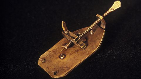
Components of light microscopes
Calculating the magnification of light microscopes
The compound microscope uses two lenses to magnify the specimen: the eyepiece and an objective lens.
In most microscopes, there is a choice of objectives to use. Magnification can therefore be varied, according to the size of the specimen to be viewed and the level of detail required.
The magnification of a lens is shown by a multiplication sign followed by the amount the lens magnifies. So a lens magnifying ten times would be ×10. The total magnification of a microscope is:
So, if the magnification of an eyepiece is ×10 and the objective is ×4, the magnification of the microscope is:
Magnification of eyepiece × magnification of objective = 10 × 4 = 40
Question
If the magnification of an eyepiece is ×10 and the objective is ×40, what is the magnification of the microscope?
×400. Because 10 × 40 = 400.
Calculating the magnification of an image
Microscopes use lenses to magnify the image of a specimen so that it appears larger.
The formula to calculate magnification is:
The same formula shown in a magnification triangle:
Calculating the magnification of a cell
The real size of the cell shown above is 0.05 mm (= 50 μm).
In a book, a photomicrograph of the cell measured 100 mm.
To calculate the magnification:
\(\text{magnification} = \frac{\text{100 mm}}{\text{0.05 mm}} = 2000\)
It is important to work in the same units when calculating magnification.
Sizes of most cells are given in micrometres, symbol μm.
Calculating the magnification, working in micrometres:
First convert the 100 mm into micrometres:
1 mm = 1000 μm
100 mm = 100 × 1000 μm = 100 000 μm
The magnification of the image calculated in μm:
\(\text{magnification} = \frac{\text{100 000 mm}}{\text{50 μm}} = 2000\)